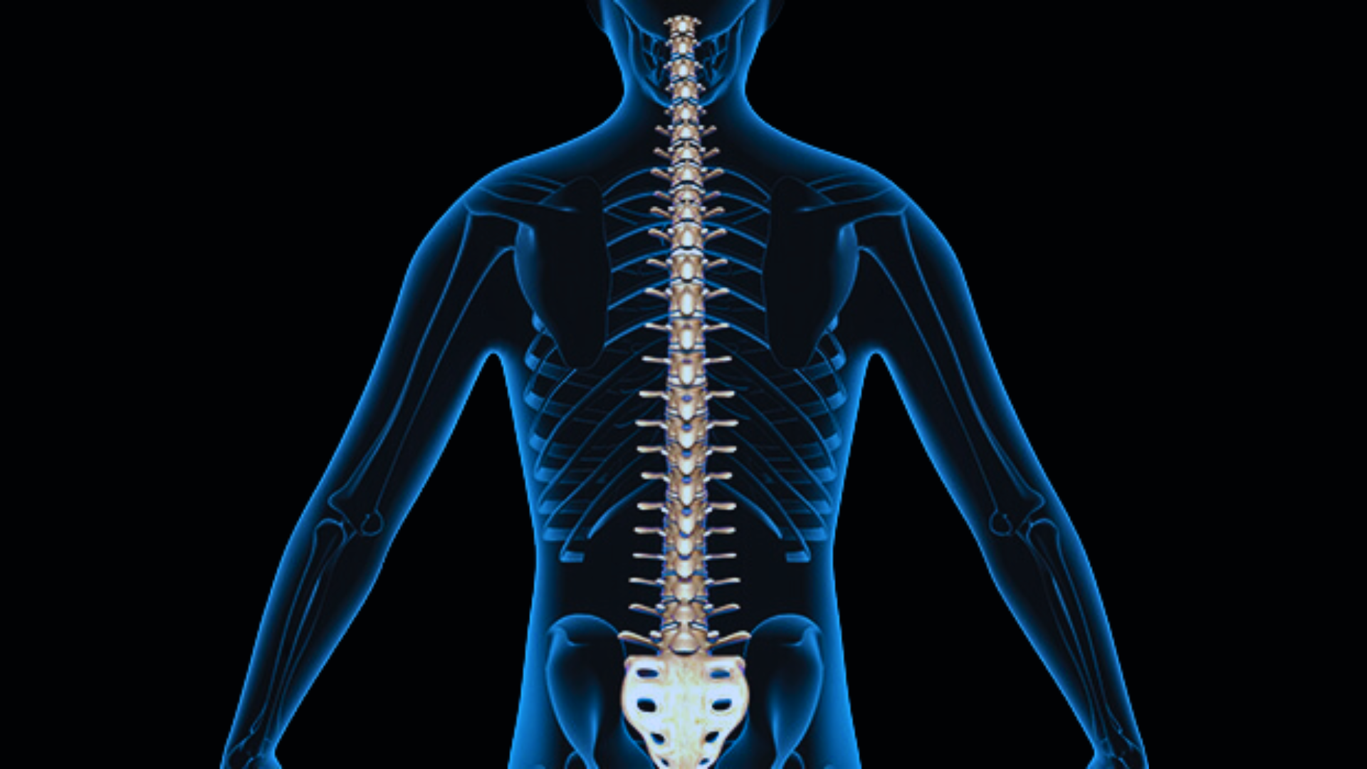
Spine
Spine Problems
Most spinal problems involving the cervical spine, thoracic spine, and or lumbar spine can be treated conservatively without surgery. Time, rest, anti-inflammatory medications, and physical therapy, are often safe and effective in the treatment of common spinal problems. Certain problems may require surgical intervention. The following procedures are commonly used to treat spinal problems refractory to non-operative care.
Minimally Invasive Spine Surgery
Advances in surgical techniques and instrumentation have led to the emergence of the field of minimally invasive spine surgery. We perform outpatient cervical discectomy and outpatient lumbar discectomy with quick patient recovery and return to daily activities. Minimally invasive lumbar decompression surgeries for spinal stenosis have also reduced length of hospital stay and have led to expedient patient recovery.
Minimally Invasive Spine Surgery
Lumbar Spine
Cervical Spine Anterior Cervical Discectomy and Fusion (ACDF)
ACDF is a common surgical procedure to treat nerve root and/or spinal cord compression secondary to disc herniation or bone spur formation. This procedure is also used to repair cervical spine injury secondary to trauma. This procedure is used when other non-surgical treatments have failed.
A cervical disc may be injured/damaged or may degenerate which may cause spinal cord or nerve root compression. This nerve root may become inflamed and cause pain, and neurological dysfunction including weakness and numbness.
After general anesthesia, a small incision is made on the anterior surface of the right or left side of the neck in order to reach the spine. The damaged disc is identified with x-ray guidance, and is subsequently removed in order to decompress the spinal cord and nerve roots. Arthritic bone spurs are also removed. The intervertebral foramen, the bone channel through which the spinal nerve exits, is then enlarged with a small instrument giving the nerve more room to exit the spinal canal.
To prevent the vertebrae from collapsing and to increase stability, the open space is often filled with bone graft, or PEEK spacer. The process of the bone graft joining the vertebrae together is called “fusion”. Sometimes a titanium plate and or screws are utilized to increase stability during fusion.
The surgery may be performed on an outpatient basis or with a 1-2 day hospital stay depending upon surgeon and patient preferences and requirements. Generally a patient may return to office work in 2-6 weeks, individual restrictions may vary.
Cervical Laminoplasty
The lamina is a flat portion of bone that is the back portion of the vertebra. When the spinal canal has become too small due to injury or disease, the canal may be made larger by use of laminoplasty. This procedure helps to alleviate spinal cord compression. An incision is made down the back of the neck to expose the cervical vertebrae. On one side of the vertebral column, the lamina are partially cut to create a hinge-like movement, much like a door. The lamina on the other side are cut all the way through to, in effect, open the “door.” After gently opening the “door” of each vertebra to create more room for the spinal cord and nerve roots behind it, bone wedges and/or titanium plates are inserted to keep the spinal canal open.
Posterior Cervical Microforaminotomy
Often referred to as “Laser Surgery,” this procedure has been available for over 60 years, but has been made less invasive through the use of finer instruments, techniques and the operating microscope. Posterior cervical microforaminoty is for select patients who present with isolated compression of nerve roots exiting the spinal cord in the cervical spine. The technique of posterior microforaminotomy allows for limited and yet effective decompression of nerve roots without committing the patient to a fusion operation. This type of operation is performed from the back of the neck and is associated with significantly less structural modifications of the spine which in the long run lessens the likelihood of accelerated degenerative disc disease and bone spur formation in adjacent levels of the spine. Potential candidates for this surgery need to undergo a thorough neurological evaluation, as not every patient is an appropriate candidate for this type of operation.
Cervical Disc Replacement with Mobi-C disc replacement
This procedure is similar to the anterior cervical discectomy and fusion (ACDF), but no fusion is performed. The damaged disc and offending bone spurs are removed through an anterior approach in order to alleviate pressure on the spinal cord and nerve roots. A synthetic disc is then inserted into the remaining space between the vertebral bodies previously occupied by the damaged disc. The goal of the intervertebral disc prosthesis, is to restore the normal dynamic function of the spine and to significantly reduce pain. This is achieved through the re-establishment of the disc height, as maintained by the prosthesis, which reduces nerve compression. Prior to the development of artificial discs the main surgical option involved fusion, in which adjacent vertebral bodies are “fused together” permanently using implants, bone chips and/or cages. The goal of the intervertebral disc prosthesis is to maintain mobility at the affected intervertebral disc and to reduce the extra loading on the adjacent intervertebral discs. This has been shown to reduce adjacent level degeneration as compared to a cervical fusion.
Product Overview
The Mobi-C® Cervical Disc (Mobi-C) has been designed for cervical disc replacement to restore segmental motion and disc height. The components of Mobi-C include superior and inferior Cobalt Chromium alloy endplates coated with plasma sprayed Titanium and hydroxyapatite coating, and a polyethylene mobile bearing insert. The controlled mobility of the patented mobile core is the foundation of Mobi-C, encouraging height restoration and respect of the instantaneous axis of rotation for a return to physiological mobility of the spinal segment. An Investigational Device Exemption (IDE) study of Mobi-C for both one and two-level cervical intervertebral disc replacement has been completed in the United States. Mobi-C is the first cervical disc FDA approved for one and two-level indications.
Cervical Disc Replacement with Prodisc C disc replacement
This procedure is similar to the anterior cervical discectomy and fusion (ACDF), but no fusion is performed. The damaged disc and offending bone spurs are removed through an anterior approach in order to alleviate pressure on the spinal cord and nerve roots. A synthetic disc is then inserted into the remaining space between the vertebral bodies previously occupied by the damaged disc. The goal of the intervertebral disc prosthesis, is to restore the normal dynamic function of the spine and to significantly reduce pain. This is achieved through the re-establishment of the disc height, as maintained by the prosthesis, which reduces nerve compression. Prior to the development of artificial discs the main surgical option involved fusion, in which adjacent vertebral bodies are “fused together” permanently using implants, bone chips and/or cages. The goal of the intervertebral disc prosthesis is to maintain mobility at the affected intervertebral disc and to reduce the extra loading on the adjacent intervertebral discs. This has been shown to reduce adjacent level degeneration as compared to a cervical fusion.
Lumbar Spine Microdiscectomy
A microdiscectomy removes a disc herniation (herniated disc) to relieve pressure on an adjoining nerve. This procedure allows for shorter hospital stays (mostly outpatient), smaller scars – 18 mm, quicker return to work and normal activities, and less post-operative pain – no muscle cutting.
Who Can Benefit
Kyphoplasty and Vertebroplasty
Kyphoplasty and Vertebroplasty are effective treatment options for patients who are suffering from intractable back pain caused by osteoporotic and pathological compression fractures of the spine. For those patients who meet the surgical criteria, these methods of stabilizing the vertebral fractures of the spine have resulted in rapid and significant reduction in incapacitating pain. Potential candidates for this operation must undergo careful screening and diagnostic work-up before surgery. The operation is generally performed on an outpatient basis.
Lumbar Laminectomy
Laminectomy is a spine operation to remove a portion of the vertebral bone called the lamina. There are many variations of laminectomy, including laminotomies, hemilaminotomies, hemilaminectomies, complete laminectomy. Generally, we prefer to remove the least amount of bone possible to accomplish the task of decompressing the spinal cord or nerve roots. This procedure was one of the first type of spinal surgeries performed, but has been refined over the years and remains quite effective in alleviating spinal cord and nerve compression.
Lumbar Spinal Fusion
In cases of spinal instability secondary to degeneration, spondylolisthesis, trauma or tumors, a lumbar decompressive procedure such as a laminectomy may be combined with a fusion procedure. Spinal fusion is a surgical technique used to join two or more vertebrae. Supplementary bone tissue, either from the patient (autograft) or a donor (allograft), is used in conjunction with the body’s natural bone growth (osteoblastic) processes to fuse the vertebrae. This procedure is used primarily to eliminate the pain caused by abnormal motion of the vertebrae by immobilizing the vertebrae themselves. There are two main types of lumbar spinal fusion, which may be used in conjunction with each other:
Posterolateral fusion places the bone graft between the transverse processes in the back of the spine. These vertebrae are then fixed in place with screws and/or wire through the pedicles of each vertebra attaching to a metal rod on each side of the vertebrae.
Interbody fusion places the bone graft between the vertebra in the area usually occupied by the intervertebral disc. In preparation for the spinal fusion, the disc is removed entirely, for example in ACDF. A device may be placed between the vertebra to maintain spine alignment and disc height. The intervertebral device may be made from either plastic or titanium. The fusion then occurs between the endplates of the vertebrae. Using both types of fusion is known as 360-degree fusion. Fusion rates are higher with interbody fusion. Three types of interbody fusion are:
In most cases, the fusion is augmented by a process called fixation, meaning the placement of metallic screws (pedicle screws often made from titanium), rods or plates, or cages to stabilize the vertebra to facilitate bone fusion. The fusion process typically takes 6–12 months after surgery. During this time external bracing (orthotics) may be required. External factors such as smoking, osteoporosis, certain medications, and heavy activity can prolong or even prevent the fusion process. If fusion does not occur, patients may require reoperation.
Some newer technologies are being introduced which avoid fusion and preserve spinal motion. Such procedures, such as artificial disc replacement, are being offered as alternatives to fusion, but have not yet been adopted on a widespread basis in the US. Their advantage over fusion has not been well established. Minimally invasive techniques have also been introduced to reduce complications and recovery time for lumbar spinal fusion.
X-LIF and ILIF
X-LIF and ILIF are two different types of Lumbar Instrumented Fusion developed to overcome the potential shortcomings of standard treatments for lumbar spinal stenosis treatments, using a minimally disruptive surgical technique. To address stenosis, ILIF involves a minimally disruptive decompression procedure called a distraction laminoplasty which involves temporary distraction of the space between the spinous processes, and careful removal of only small sections of bone to relieve the pressure on the spinal cord and nerves. During ILIF, a precision-machined allograft bone is placed between the spinous processes to permanently distract areas that are pressing on the spinal cord and/or nerves, promote fusion between the spinous processes to provide long-term spine stabilization, and provide a protective cover for the spinal cord to help prevent scar tissue from pressing on the spinal cord and/or nerves. A small plate is then attached to both spinous processes to stabilize the segment of the spine and promote fusion, eliminating the need for more extensive surgery.
The XLIF® Surgical Procedure
After you have been positioned, an X-ray will be taken to help your doctor precisely locate the operative space. Next, your skin will be marked at the site where the two small incisions will be made. Your surgeon will use the latest instrumentation to access the spine in a minimally disruptive manner. Disc preparation is the next step. This is done by removing the disc tissue which will allow the bones to be fused together. Several X-rays will be taken during this stage to ensure the preparation is correct. Once the disc has been prepared, the surgeon will then place a stabilizing implant into the space to restore the disc height and enable the spine to once again support necessary loads. Once in position, a final X-ray will be taken to confirm correct implant placement. In the event that further stabilization is necessary, the surgeon may choose to insert additional screws, rods, or plates into the vertebrae.
Complex Spine Surgery
Anterior and posterior, cervical, thoracic, and lumbosacral instrumented spinal fusion operations are examples of complex spine surgery that we perform on a regular basis. Spinal fusion operations are procedures of choice for a carefully selected sub-group of patients who have progressive and medically intractable back pain. Spinal fusion and instrumentation operations are often effective treatment modalities for spinal instability caused by degenerative processes, spinal infections, tumors and severe trauma to the spine. We have extensive experience and training in complex spinal fusion and instrumentation and treat a large number of patients with these forms of spinal disorders.
Lumbar Disc Replacement with Synthes Prodisc L
With the introduction of total disc replacement (TDR) surgery, surgeons can offer their patients an alternative to spinal fusion surgery for the treatment of symptomatic degenerative disc disease (DDD). The TDR procedure is intended to relieve pain and preserve motion in the spine.
During both TDR surgery and spinal fusion surgery, the pain-generating disc is removed and the normal disc height is restored. During a fusion surgery, the spinal segment is stabilized with an implant and plate and/or rods and screws. Bone graft may be used to promote osseous fusion of the vertebrae. Conversely, during a TDR surgery an implant that allows motion is inserted into the disc space.
Both treatments are usually effective for relieving pain. However, preserving motion at the treated vertebral segment may enable the spine to restore its sagittal balance and maintain more natural mechanics after surgery than fusing the vertebral segment. This may potentially decelerate degeneration in healthy adjacent levels in the spine. Link to Synthes Pro Disc L.